Beranda
/ Anatomy Of Chest And Ribs : Chest Wall Amboss / The other end is blunt and smooth.
Anatomy Of Chest And Ribs : Chest Wall Amboss / The other end is blunt and smooth.
Insurance Gas/Electricity Loans Mortgage Attorney Lawyer Donate Conference Call Degree Credit Treatment Software Classes Recovery Trading Rehab Hosting Transfer Cord Blood Claim compensation mesothelioma mesothelioma attorney Houston car accident lawyer moreno valley can you sue a doctor for wrong diagnosis doctorate in security top online doctoral programs in business educational leadership doctoral programs online car accident doctor atlanta car accident doctor atlanta accident attorney rancho Cucamonga truck accident attorney san Antonio ONLINE BUSINESS DEGREE PROGRAMS ACCREDITED online accredited psychology degree masters degree in human resources online public administration masters degree online bitcoin merchant account bitcoin merchant services compare car insurance auto insurance troy mi seo explanation digital marketing degree floridaseo company fitness showrooms stamfordct how to work more efficiently seowordpress tips meaning of seo what is an seo what does an seo do what seo stands for best seotips google seo advice seo steps, The secure cloud-based platform for smart service delivery. Safelink is used by legal, professional and financial services to protect sensitive information, accelerate business processes and increase productivity. Use Safelink to collaborate securely with clients, colleagues and external parties. Safelink has a menu of workspace types with advanced features for dispute resolution, running deals and customised client portal creation. All data is encrypted (at rest and in transit and you retain your own encryption keys. Our titan security framework ensures your data is secure and you even have the option to choose your own data location from Channel Islands, London (UK), Dublin (EU), Australia.
Anatomy Of Chest And Ribs : Chest Wall Amboss / The other end is blunt and smooth.. The rib cage is a bony structure found in the chest (thoracic cavity). The rib cage also anchors the bones of the head, neck, shoulders, and arms to the trunk of the body. Flail chest describes what happens when blunt force trauma to the chest causes multiple adjacent ribs to fracture and separate from the chest wall. Learn about each of these muscles, their locations, functional anatomy and exercises for them. Illustration of human body anatomy from antique french art book:
Note the big rib sign on the lateral radiograph (larger more posterior appearing ribs are the right ribs. Contributing to their role in protecting the internal thoracic organs. The bones of the chest and upper back combine to form the strong, protective rib cage around the vital thoracic organs such as the heart and lungs. It is made up of 12 pairs of ribs. Flail chest describes what happens when blunt force trauma to the chest causes multiple adjacent ribs to fracture and separate from the chest wall.
Thoracic Wall And Breast Illustrations from www.imaios.com Flail chest describes what happens when blunt force trauma to the chest causes multiple adjacent ribs to fracture and separate from the chest wall. The bones of the chest and upper back combine to form the strong, protective rib cage around the vital thoracic organs such as the heart and lungs. Rib cage, in vertebrate anatomy, basketlike skeletal structure that forms the chest, or thorax, and is made up of the ribs and their corresponding attachments to the sternum (breastbone) and the vertebral column. It discusses the specific anatomy of the ribs and costal cartilages, along with the sternum. The cartilage strips are called costal cartilage (costal is the anatomical adjective that refers to the rib) and connect on their other end to the sternum. In humans and other hominids, the thorax is the chest region of the body between the neck and the abdomen, along with its internal organs and other contents. The palpable midline sternum is variable in size and shape; The first seven ribs progressively increase in length, the lower five ribs then begin to decrease in length.
At the chest, many rib bones connect to the sternum via costal cartilage,.
The palpable midline sternum is variable in size and shape; They articulate with the vertebral column posteriorly, and terminate anteriorly as cartilage (known as costal cartilage). The chest wall is a complex system that provides rigid protection to the vital organs such as the heart, lungs, and liver; The 12th rib is not always present. Anatomynote.com found chest bone, ribs, lung, heart, xiphoid process, sternum anatomy from plenty of anatomical pictures on the internet. Flail chest describes what happens when blunt force trauma to the chest causes multiple adjacent ribs to fracture and separate from the chest wall. The bones of the chest and upper back combine to form the strong, protective rib cage around the vital thoracic organs such as the heart and lungs. The other end is blunt and smooth. Powerful muscles that move the head and arms attach to these bones as well. There are twelve pairs of ribs, all of which articulate with the vertebral column. Ribs are highly vascular and trabecular with a thin outer layer of compact bone. There are 12 pairs of ribs which are separated by intercostal spaces. The rib cage also anchors the bones of the head, neck, shoulders, and arms to the trunk of the body.
There are twelve pairs of ribs, all of which articulate with the vertebral column. The rib cage also anchors the bones of the head, neck, shoulders, and arms to the trunk of the body. This image added by admin. Both the liver and the stomach are located in the lower chest region under the thoracic diaphragm, a sheet of muscle at the bottom of the rib cage that separates the chest cavity from the abdominal. For more anatomy content please follow us and visit our website:
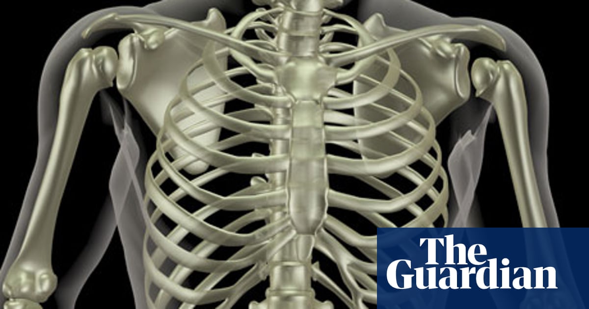
Mapping The Body Ribs Human Biology The Guardian from i.guim.co.uk It is made up of the manubrium superiorly, the body and the xiphisternum (figure 1).the manubrium has an upper central depression, the suprasternal notch. Powerful muscles that move the head and arms attach to these bones as well. Rib cage, in vertebrate anatomy, basketlike skeletal structure that forms the chest, or thorax, and is made up of the ribs and their corresponding attachments to the sternum (breastbone) and the vertebral column. Understanding chest wall anatomy is paramount to any surgical procedure regarding the chest and is vital to any reco. It discusses the specific anatomy of the ribs and costal cartilages, along with the sternum. We hope this picture chest bone, ribs, lung, heart, xiphoid process, sternum anatomy can help you study and research. The chest wall is a complex system that provides rigid protection to the vital organs such as the heart, lungs, and liver; Ninja nerds!join us in this video where we show the sternum and rib articulation anatomy through the use of a model.
Find out more about the individual muscles within the chest anatomy by clicking their respective links throughout this
On an individual rib, one end has various processes, facets, and bumps. They are extremely light, but highly resilient; The ribcage contains 12 ribs total on each side, divided into three different types. Ribs are highly vascular and trabecular with a thin outer layer of compact bone. The posterior rib (right) is farther from the film and is magnified more than the anterior rib (left), which is in contact with the film. Chest anatomy the chest, also called the thorax, contains several key anatomical structures and organs. Rib cage, in vertebrate anatomy, basketlike skeletal structure that forms the chest, or thorax, and is made up of the ribs and their corresponding attachments to the sternum (breastbone) and the vertebral column. Each pair is numbered based on their attachment to the sternum, a bony process at the front of the rib cage which serves as an anchor point. The chest anatomy includes the pectoralis major, pectoralis minor and the serratus anterior. The chest wall is formed from the sternum anteriorly, 12 pairs of ribs, costal cartilages and intercostal muscles laterally, and the thoracic vertebrae posteriorly. You can click the image to magnify if you cannot see clearly. 1 the ribcage protects some of the most important organs, including the lungs and the heart. This is the end that articulates with the vertebra.
They articulate with the vertebral column posteriorly, and terminate anteriorly as cartilage (known as costal cartilage). However, only seven have a direct articulation with the sternum. The top seven ribs (called the true ribs) connect with cartilage to the breastbone (sternum). We hope you can get the exact information you. Note the big rib sign on the lateral radiograph (larger more posterior appearing ribs are the right ribs.
Rib Cage Anatomy Function Britannica from cdn.britannica.com It is mostly protected and supported by the rib cage, spine, and shoulder girdle. Find out more about the individual muscles within the chest anatomy by clicking their respective links throughout this Contributing to their role in protecting the internal thoracic organs. For more anatomy content please follow us and visit our website: The ribs protect and make space for the heart, lungs and other organs of the chest and abdomen. We think this is the most useful anatomy picture that you need. The bones of the chest and upper back combine to form the strong, protective rib cage around the vital thoracic organs such as the heart and lungs. The chest wall is formed from the sternum anteriorly, 12 pairs of ribs, costal cartilages and intercostal muscles laterally, and the thoracic vertebrae posteriorly.
However, only seven have a direct articulation with the sternum.
Contributing to their role in protecting the internal thoracic organs. Chest anatomy the chest, also called the thorax, contains several key anatomical structures and organs. And flexibility to aid in the functional process of respiration. The rib cage also anchors the bones of the head, neck, shoulders, and arms to the trunk of the body. The ribs protect and make space for the heart, lungs and other organs of the chest and abdomen. Note the big rib sign on the lateral radiograph (larger more posterior appearing ribs are the right ribs. The first seven ribs progressively increase in length, the lower five ribs then begin to decrease in length. The other end is blunt and smooth. The anatomy of the human ribs (costae) are one of the integral parts of the chest wall; Stability to arm and shoulder movement; This image added by admin. Powerful muscles that move the head and arms attach to these bones as well. We think this is the most useful anatomy picture that you need.
The top seven ribs (called the true ribs) connect with cartilage to the breastbone (sternum) anatomy of chest. Thank you for visit anatomynote.com.